Friday, May 20, 2016

Cardio Lab

The next step in understanding the cardiovascular system is to measure how the heart physically reacts to certain stimulus (i.e. emotion, exercise, food intake, etc.). To accomplish this, we were asked to design a lab in which we tested subjects with one of these stimuluses while monitoring their heart activity.

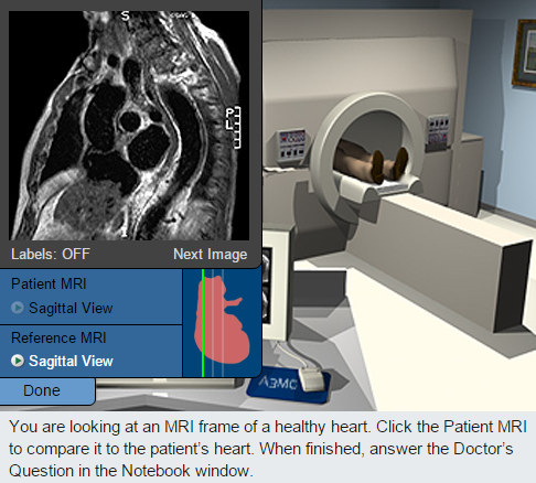
My lab partner and I chose to take advantage of the election year by using videos of politicians as an emotional stimulus in a blood pressure monitoring experiment. To begin the lab process, we gathered five volunteer participants. With each test subject, we conducted a survey to determine their basic political views. The survey is as follows:

1) Which candidate is your first choice for the 2016 election? (Donald Trump, Ted Cruz, Hillary Clinton, or Bernie Sanders)

2) Which candidate is your last choice for the 2016 election? (Donald Trump, Ted Cruz, Hillary Clinton, or Bernie Sanders)

3) Which political party do you most agree with? (Democratic, Republican, or Independent)

4) What information are you aware of regarding Hillary Clinton's involvement in the Benghazi events that occurred on September 11, 2012, as well as her Benghazi hearings?

5) What information are you aware of regarding the recent Planned Parenthood events?

6) What information are you aware of regarding the government shutdown, as well as Ted Cruz's involvement in the situation?

7) What information are you aware of regarding Donald Trump's "wall" policy proposal?

8) How would you define socialism? Does socialism contradict or enforce American values (the ideals put in place by the Founding Fathers)?


The results from the survey are shown here:
Subject A
Supported Bernie Sanders the most, Donald Trump the least. (Democrat)

Subject J
Supported Bernie Sanders the most, Donald Trump the least. (Independent)

Subject T
Supported Bernie Sanders the most, Donald Trump the least. (Democrat)

Subject D
Supported Ted Cruz the most, Bernie Sanders the least. (Republican)

Subject B
Supported Ted Cruz the most, Hillary Clinton the least. (Republican)


After completing the survey, we evaluated their answers and selected videos according to their responses. We showed them both the pros and cons for each candidate they chose (first choice and last choice). To measure their heart activity, we took their blood pressure before and after showing them the pros and cons for each candidate.  The videos we showed are linked below:

Hillary Clinton Con:
https://www.youtube.com/watch?v=8BfNqhV5hg4

https://www.youtube.com/watch?v=BQ7qfnaYSTA&ebc=ANyPxKp1uu5-4-MNKm_-oraSvgsFRDaSvj-P_vYd-qaaGFvt5wLkOGMtQtP_X5porvL2ipErT99XeTRohuJ2mXUbBgNAVR684g

Ted Cruz Con:
https://www.youtube.com/watch?v=bZC8VPz-ySM

http://youtu.be/EOBq2TatJ0M

Donald Trump Con:
https://www.youtube.com/watch?v=ptUpK_y4g8k

Bernie Sanders Con:
https://www.youtube.com/watch?v=3g7cz5aXSsQ


Bernie Sanders Pro:
http://youtu.be/nyhfJTFJHu8

Before we began the experiment, my lab partner and I hypothesized that if we exposed several people to videos that contradicted their ideas, a negative emotional response would be elicited, resulting in an increase in their blood pressure.

The results from the lab are shown below:


Once this data was collected, we complied the information and found the average percent of change in blood pressure. This data was then organized in the following graph:


After reviewing the data, we observed that the participants' reactions to the experiment were highly varied and did not carry a distinct pattern of reaction. Therefore, the data did not support a distinct conclusion for the lab.

In order to communicate these results to the public and scientific community, my lab partner and I created a poster with the information from the lab and displayed it in the hallway. This poster is pictured below.


Although the results from this lab were slightly disappointing, I found this project to be very beneficial in aiding my understanding of blood pressure and heart activity. I hope you enjoyed it as much as I did!


Friday, March 11, 2016

Virtual Cardiology Lab

As we look closer into the cardiovascular system, we completed an online lab to learn about how symptoms for heart disease can be detected, as well as the tools and techniques that are used to detect such symptoms.

In this virtual lab, we had the ability to step into the shoes of a medical intern, testing three different patients who believed they might have a heart condition. To confirm/deny their suspicions, each patient was put through three analysis examinations: stethoscopic, echocardiographic, and MRI. Their pedigrees were also analyzed to determine whether or not, if they did have a heart condition, the condition was dominate or recessive. As we went through the online lab, a series of reference photos were given to demonstrate what a normal heart should look and sound like during these tests; these were beneficial in the process of determining whether or not the patient had a condition, and what, specifically, might be the issue.

Reference Photos 


Patient A
The first patient we examined was patient A, a 30 year old male of tall stature and light weight. His records can be seen below. 


After reviewing patient A's file, he was taken into the stethoscopic room for an exam. 


After conducting the exam, we were able to compare the patient's heart sound to the healthy reference heart sound, and we found them to be very different. The healthy heart had a very distinct and separated sound while the patient's sound was more warbled, blending together in some places and delayed in others. This sound indicated a systolic murmur, and the patient was encouraged to undergo more testing. 

The next test performed was an echocardiographic exam, in which it was concluded that the patient's aorta was increased in size, indicating the presence of an aneurysm. The results showed that further testing would be required.


The next test performed was an MRI exam. After comparing the test results to the reference MRI, it became clear that the patient was suffering from a dilated aorta, which is a form of an aortic aneurysm.

 As the patient mentioned earlier, their sibling died of heart related causes. After evaluating patient A, we reviewed his pedigree to determine whether or not the condition was inheritable, and whether or not any other family members were at risk.

 A quick evaluation of the pedigree confirmed that there was a dominant pattern of inheritance. 


All of the results from the tests and examinations were considered, leading to a diagnosis of Marfan Syndrome. 


Patient B

The next patient examined was a 35 year old male, with a dwarfic condition.


Just like with patient A, patient B's examination began with a stethoscopic test. Again, the sound heard was abnormal, with the "lub" and "dub" being heard as multiple short sounds than singular, distinct noises. 


The next test was the echocardiographic exam. The results showed a very distinct issue: the patent was missing the wall between the left and right atria, a severe form of an atrial septal defect. This condition could lead to a flooding of blood in the lungs as well as an imbalance of pressure in the heart.


The patient's pedigree was then examined, and it was found that the condition could be inherited through a recessive gene.


The results from the patient examination lead to a conclusive diagnosis of Ellis-van Creveld (ECV) syndrome. This condition is a rare and recessive gene characterized by atrial septal defects as well as dwarfism, which are both seen in the patient.


Patient C

The last patient examined was a 55 year old male of normal build. 

To gain an understanding of the patient's condition, a stethescopic exam was conducted. A murmur could be heard between the second and first sound, which prompted further testing.


The next test was the echocardiograph. A comparison between the echocardiograph and the reference echocardiograph indicated that the patient had a thickened leaflet in the mitral valve, causing it to move slowly and poorly. This has led to an obstruction between the left atrium and left ventricle; the patient is suffering from a condition known as mitral stenosis. Since the condition has been identified, it is not necessary to take an MRI exam, however, the pedigree still needs to be evaluated.


Since only members of the patient's generation have the condition, it is likely not inherited dominantly, and since there are so many people with the condition, it is most likely not inherited recessively either. It is most likely that this condition is a result of environmental factors. 


Here is some more information on mitral stenosis and how it relates to the patient:


Cardiovascular Physiology: Monitoring the Heart

To further our understanding of the cardiovascular system, we began to explore the physiology of the system by conducting another EKG lab.

In this lab, it was our goal to collect an EKG reading of a pulse and evaluate the P,Q,R,S, and T wave intervals, as well as determine the heart rate (beats per minute). To do this, we used and EKG sensor that was connected to the computer and attached to the subject's arm by three electrode tabs. The green (negative) and black (ground) tabs were attached to the right arm, and the red (positive) tab was attached to the left arm.

As the data was collected, the subject sat with their arms resting on their legs, allowing an accurate collection of data. After a few moments, the computer presented a data chart:


To fully understand this lab, it is important to know the different waves in a pulse. The waves have 5 main categories: P, Q, R, S, and T. The P wave is the first wave to occur, and it helps the heart "gear up" for the action part of the beat, which occurs through waves Q, R, and S.  Recovery from the beat occurs during wave T. 


After the pulse was recorded, the intervals between each wave was measured, and the heart rate was calculated. We compiled this data into another chart:


As you can see, some of the data we collected differed from the "Standard Resting Electrocardiogram Interval Times". This is most likely because we conducted the lab with the electrode tabs on the arms rather than directly over the heart, which caused a slight variation in the accuracy of the data.  Overall, the data was fairly close to what it was expected to be. 

During this lab, we were able to visualize and measure cardiovascular activity, as well as recognize the different wave forms seen in an EKG reading. Through this, our understanding of cardiovascular activity was enhanced. 

Heart Anatomy

As we wrap up the neurophysiology unit, we are beginning to switch focus to the anatomy and physiology of the cardiovascular system. To kick things off, we dissected a pig heart, sheep heart, and cow heart, measuring and comparing the main components. Before we began the dissection, we filled out a heart diagram to become familiarized with heart anatomy.


During the dissection, we took measurements of the hearts so that we would be able to compare not only part to part, but heart to heart as well. These measurements are shown in the chart below, followed by pictures of the measurements being taken.


Sheep Heart


Pig Heart


Cow Heart


I hope you enjoyed this post; I found it very interesting to explore the cardiovascular anatomy!