Tuesday, November 3, 2015

Integumentary System

In this blog post, we are going to discuss the normal structure of skin and how certain conditions can change the normal structure and functions of the skin. Specifically, we are going to look at tattoos, laser hair removal, and poison ivy. Before we get into that, however, we should take a look at how skin functions under normal conditions.

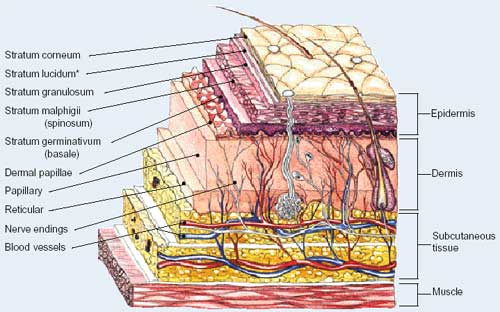
The skin consists of three basic layers: the epidermis, dermis, and hypodermis. Within these layers, there are other layers as well. The epidermis is composed of five layers: stratum basale (basale layer), stratum spinosum (prickly layer), stratum granulosum (granular layer), stratum lucidum (clear layer), and stratum corneum (horny layer). These five layers contain four distinct cell types, including melanocytes, Merkel cells, Langerhan's cells, and keratinocytes. The dermis consists of two layers: the papillary and reticular layers. There are other components to the skin as well, such as sweat glands, blood vessels, and hair follicles. The skin layers, under normal conditions, are shown below.


The picture below illustrates the process of dying the skin through tattoos. The needle pierces the epidermis and injects ink into the dermis in the papillary layer. The needle is motorized, puncturing the skin between 50 and 3,000 times per minute, depositing ink with every puncture. The ink can then be seen through the epidermis, and due to the stability of the cells in the dermis, the ink does not travel or fade in the skin. 


In the next picture, the integumentary system's reaction to poison ivy is displayed. An irritant from the ivy is transferred to the epidermis where it immediately triggers a reaction. Effector T cells signal white blood cells to attack the site of intrusion. A water soluble, nonallergenic compound is then formed, which prevents further allergic response. The Effector T cells then stop signaling the white blood cells, and natural antiseptic properties sanitize the rash, which is healed within 3 days. 


The last condition we are going to look at is electrolysis, or laser hair removal. In this case, a needle is inserted into the skin to come in contact with a hair follicle. An electrical shock is transmitted to the base of the follicle, causing it to reach a temperature in which hair growth is irreversibly inhibited. The epidermis, located below, remains undamaged.

http://www.follikill.com/wp-content/uploads/2014/01/how-laser-hair-removal-work1.jpg


I hope you enjoyed learning about the integumentary system!

Wednesday, October 7, 2015

Human Models of Epithelia


Epithelial tissues are composed almost entirely of cells, and are supported by connective tissue. Within epithelial tissues, there are orginizational categories including: simple and stratified. Within those categories, there are also three types: squamous, cuboidal, and columnar.  In this post, our classmates worked together to create human models of the aforementioned epithelial tissues. 

The first model we made was for pseudostratified columnar epithelial tissue. This is a single layer of cells, in which all of the cells are of varying heights. Some cells do not reach the surface, and the nuclei can be seen on different levels. This type of epithelial tissue secretes mucus and propels it through ciliary action. Pseudostratified columnar epithelial tissue can be found lining the trachea and most of the upper respiratory tract. Nonciliated forms of this tissue can be found in male's super-carrying ducts and ducts of large glands.


Transitional epithelial tissue consist of many cell layers in which the cells located at the base are cuboidal and the surface cells are shaped like domes. Translational epithelia can be found lining the urinary bladder, ureters, and part of the urethra, stretching to permit the distension of the urinary bladder. 

The simple squamous is a stout, squished layer of single cells that diffuses and filters. It is used in the lymphatic and cardiovascular systems, creating a thin, slick layer that reduces friction. 


Stratified cuboidal is more uncommon in the body, and is usually 2-3 layers thick. Stratified cuboidal tissue can be found in some mammary and sweat glands.


Simple cuboidal epithelia is another example of epithelia that absorbs and secretes. This type of epithelia is found in the kidney tubules, ovary surface, and ducts and secretory portions of small glands. Simple cuboidal epithelial tissue is composed of a single layer of cube like cells with large, spherical central nuclei. 


Simple columnar epithelia consist of a single layer of tall cells with oval nuclei. Many of these cells contain cilia, which help transport mucus. This layer may also contain mucus-secreting unicellular glands referred to as goblet cells. Located in the digestive tract, gallbladder, and excretory ducts of some glands, nonciliated simple columnar epithelia works to absorb, secrete mucus, enzymes, and other substances. Ciliated simple columnar epithelia is found lining small bronchi, uterine tubes, and some regions of the uterus, working to propel mucus.

Stratified columnar tissue is also not very abundant in the body, however, it can be found in the pharynx, male urethra, lining of some glandular ducts, and transitional areas between 2 types of epithelia.


And thus concludes the lesson on epithelia! Thanks to my classmates and their efforts in making this lesson!

Histology Microscope Lab


When discussing tissues, there are four broad categories: epithelial, connective, muscle, and nerve. For this post, we are going to use a microscope to view these types of tissue on a much greater scale, describing each as we work. All of the photos were taken during the lab.

The first sample we observed was that of hyaline cartilage, which is classified as a connective tissue. When functioning, hyaline cartilage supports, reinforces, cushions, and resists compressive stress.
Hyaline cartilage is composed of a network of collagen fibers that have formed an imperceptible network and chondroblasts that produce the matrix.
   

The next sample we worked with was bone, also a type of connective tissue. Bone is comprised of hard, calcified matrix that contains many collagen fibers.    Bone works to support and protect while providing leverage for the muscles. In addition, bone stores calcium, minerals, and fat, and bone marrow is home to cell formation.


Next, we looked at smooth muscle tissue. Spindle-shapes cells make up this tissue, arranging themselves closely to form sheets that propel substances or objects along passageways without voluntary control.


We also looked at cardiac muscle. When in action, contracting tissue propels blood into circulation, also without voluntary control. This tissue is composed of branching, striated, and generally uninucleate cells that interdigital with at specialized junctions.


This next sample is taken from an involuntary muscle, meaning that it originates either in the cardiac system, or as a smooth muscle tissue in the walls of hollow organs. When compared to cardiac tissue, it does not share as many characteristics as it does when compared to smooth muscle tissue. It appears to have closely arranged cells with central nuclei, as do the cells found in smooth muscle tissue.


The next two samples are those of nerve endings and neuron motors.  These are composed of branching cells, with the cell processes extended from the nucleus-containing cell body. These transmit electrical signals, controlling activity in the body.

Monday, October 5, 2015

Homeostasis Lab


In a previous post, we discussed homeostasis and how it functions. The next question to ask is: how can we prove homeostasis occurs with changes to our internal/external environment? To answer this question, our group designed a lab in which our blood glucose levels were measured as we altered our diets.

For the first part of our lab, each member in our group fasted, eating nothing and drinking only water for 24 hours. We measured our blood glucose levels before, during, and after the fast. Considering the aspects of the experiment, it was hypothesized that our blood glucose levels would decrease. This was then supported through the experimental results, which are illustrated below. 


As you can see, the blood glucose levels decreased during the 24 hours. In the first graph there is an increase in glucose levels after the 24 hours, which occurred due to the fact that the subject took their blood after eating when the fast ended, where the other subjects did not.These readings were very interesting, as a hypoglycemic and vegan were tested within the experiment. Overall, this experiment was successful in proving homeostasis.


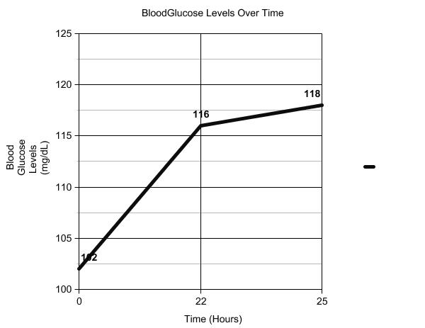
The second part of the lab involved one member of our group eating nothing but sugar-based foods for 24 hours, again measuring their blood glucose levels before, during, and after the 24 hours. For this part of the experiment, it was hypothesized that the blood glucose levels would increase over the 24 hours. The results are shown below.


As you can see, the glucose levels increased over the 24 hours, from 102 mg/dL to 118 mg/dL, again successfully proving homeostasis. 


Tuesday, September 29, 2015

Organization of the Body

This post is going to detail the different aspects of organizing the body, such as body directions, planes of section, and body cavities, while using the correct anatomical terms.


The picture above displays the anatomical position. This position requires an erect body, palms and face directed forward. This position is very common in anatomy, as it easily shows correlation between the parts of the body.

The next picture demonstrates body directions in regards to what is located above/below another body part while the body is in anatomical position. These directions are known as "Superior" and "Inferior". For an example, the head is superior to the waist while the feet are inferior to the knees.


This next picture also demonstrates direction, though this focuses more on "in front" or "behind". In anatomy, this is referred to as "Anterior" and "Posterior". The navel is anterior to the spine, and the spine is posterior to the navel.


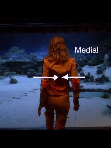
The next two images featured below illustrate two different body directions. Medial refers to body parts located toward the midline of the body while lateral refers to body parts located away from the midline of the body. The spine is medial to the shoulders while the shoulders are lateral to the spine.


Due to the mobility of appendages, there are specific directions to classify what is close/far relative to the body. The farther something gets from the trunk is described as distal while the closer something is to the trunk is described as proximal.  The fingers are distal to the elbow, and the shoulder is proximal to the wrist. 


The last directional terms we are going to discuss are superficial and deep. These reference how close  something is to the surface of the body. The spine is deeper than the skin, and the skin is more superficial than your skeleton.


Newt, we are going to go over the planes of the body. The Saggital Plane divides the "left" and "right" sides of the body.


The Transverse Plane divides the body into superior and inferior sections. 


The Frontal Plane separates anterior from posterior.


The body also has many cavities, or spaces that have the potential to be hollow. The picture below shows many of these cavities. You can even go to a smaller scale than that shown, for example, the nasal cavities, orbital cavities, etc.


Another think to look at are membranes, specifically those surrounding the heart, which are illustrated in the picture below. The visceral pericardium membrane directly covers the heart, with the parietal pericardium membrane on the outer side. The pericardial cavity divides the two, which is filled with biological fluids.


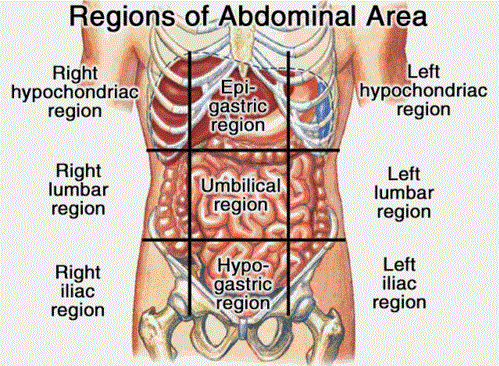
The last thing we are going to discuss in this post is the regions of the core/trunk. There are nine of these regions. They help organize the abdomen and the parts of the body associated with it. 


There you have it-the basic ways to organize the human body! 


Thursday, September 3, 2015

Let's Talk About Homeostasis

Today we are going to talk about homeostasis and figure out exactly what it is, while learning about some examples and important terms.

To begin, let's find out what homeostasis means. By definition, it is "the tendency toward a relatively stable equilibrium between interdependent elements, especially as maintained by physiological processes." So, essentially, homeostasis is where the body uses physiological processes to maintain a balance and stable environment within the body.


We are constantly putting our bodies through changing environments and altering the patterns of what we consume, but even so, our bodies manage to stay balanced through homeostasis. For example, when the environment outside of the body is cold, homeostasis makes sure the internal systems inside the body keep it at the correct temperature. 


The picture above illustrates another example of homeostasis. As you run, your body generates more heat than when you are at rest. Your body is also subjected to the weather outside, and must adapt to the temperature to maintain an internal balance. To do this, homeostasis causes increased respiratory, sweat, and radiation production. 

Another important part of homeostasis is feedback. Feedback is separated into two categories: positive and negative. 

Negative feedback- "the diminution or counteraction of an effect by its own influence on the process giving rise to it, as when a high level of a particular hormone in the blood may inhibit further secretion of that hormone, or where the result of a certain action may inhibit further performance of that action."

Negative feedback isn't a "bad" thing. Basically, something happens to your body, such as an increase in your blood glucose level, and as a result, your body has a reaction, such as secreting insulin that stores the glucose as glycogen. Because of the insulin reaction, your body begins to detect lower levels of glucose, and therefore, slow insulin secretion. This is negative feedback; your body was able to balance blood glucose levels by starting/stopping insulin production. This example is illustrated below.


Positive Feedback-"the enhancement or amplification of an effect by its own influence on the process that gives rise to it."

Positive feedback is basically where your body detects something and reacts to the change. Unlike negative feedback, the body continues to react until the disturbance is completely controlled. For example, when a woman is giving birth, the baby's head being pushed causes nerve impulses which lead to the contraction of the uterus. This pushes the baby forward, and again, a contraction occurs. this cycle repeats until the baby is pushed out. This example is also shown below. 


So there it is-the basics of homeostasis, its functions, and positive/negative feedback!