Wednesday, March 2, 2016

Sheep Brain Dissection


As the class began to learn more about nerves and the nervous system, we had the opportunity to look at a sheep's brain, and through the dissection process, we were able to understand its basic anatomy. To maximize this learning opportunity, we dissected three brains and cut them on three different planes: saggital, frontal, and transverse.  During this dissection lab, we were asked to label pictures of the brain, which can be seen below.


Monday, February 22, 2016

The Effects of Heroin on the Brain

As we dig farther into neurons and their physiology, we are beginning to look at how they function in the brain. While doing this, our class was assigned to select a research topic that would enhance our understanding of neuron functionality. For this assignment, I chose to research how heroin affects the brain. My presentation on this topic can be found here; I hope you enjoy it!

Monday, February 8, 2016

Neurophysiology


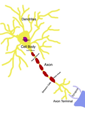
Neurophysiology is a very interesting subject that works with three main components: neurons, nervous systems, and nerves. Today, we are going to look at how they function, beginning with the anatomy of the neuron.


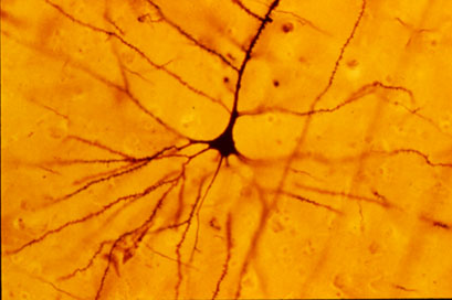
The neuron is a cell, and so it has many common components such as a nucleus, cell membrane, cell body, and different channels that ions can travel through. The neuron works to communicate signals to other neurons, muscles, and even glands. When they communicate, the signal they send causes the muscle to contact, the gland to secrete, or the next neuron to continue passing the signal through the nervous system. Neurons come in many shapes and sizes, however, all neurons have dendrites and one axon that extends from the cell body, a receptive portion, and a signaling portion.

The axon acts as the conducting or transmitting portion of the neuron, passing the signal along to the next neuron through the axon terminal and synapse. The axon connects to the cell body through the axon hillock. Axons can be from 1-2 inches to longer than a meter.

The cell body acts as a communicator between the dendrites and the axon. As the signal is received through the dendrites, the cell body passes it along to the axon. The cell body is also the main nutritional and metabolic portion of the cell.

The dendrites act as the cell receptors, receiving signals from other neurons and passing those signals through the cell. The flow of energy is directional in neurons, flowing from the dendrites, through the cell body and axon, into the next cell.

Some axons are covered in insulation bodies called myelin sheaths. These sheathes are made when Schwann cells wrap around the axon, releasing cytoplasm as it compresses in a spiral-like structure. The spaces between these sheaths are known as nodes of Ranvier. These nodes occur between every sheath, and allow ions to flow across the membrane.


There are four main channel types within the nervous system: the Sodium leak, Voltage Gated Na+, K+ Potassium Leak, and Voltage-Gated K+ channels. These ion channels are embedded in the neuron membrane, and work to control the movement of ions across the membrane. They are selective, regionally located, functionally unique, and either passive or active. Without these channels, there would be no possibility of neuron excitability. 

Ions channels are selective, in that the ions they permit to cross the membrane must fit a certain criteria. The ion must have a certain size, charge, and must attract and hold a certain level of water. These standards vary between the different channel types. 

There are also active and passive channel types. Active channels open and close, while passive channels are always open and allow ions to flow freely across the membrane. 

Some active channels have gates that are controlled by voltage; membranes with more positively concentrated ions on the outside and more negatively concentrated on the inside create membrane potential, or resting potential, producing a voltage that opens the channel. Channels can also be opened by chemical processes. Neurotransmitters can bind to certain active channels and force them open, allowing ions to travel through. 

Different ion channel types can be found on different parts of the neuron. Passive channels can be found on the dendrites, cell body, and axon; chemically gated channels can be found on the dendrites and cell body; voltage gated channels can be found on the axon hillock, unmyelinated axons, and the nodes of Ranvier. 

The Sodium/Potassium pump works to keep a balance of sodium and potassium inside and outside of the cell. The pump works with an active process involving ATP hydrolysis, transforming ATP into ADP+P, which allows potassium to enter the cell and forces sodium out. 

Resting potential, or RP, occurs when the inside of a neuron is more negative (-70mV) than outside of the neuron membrane. No change in voltage will occur over time, and the sodium and potassium will not reach equilibrium, as the outside of the neuron will have more sodium and the inside will have more potassium. 

Synaptic potentials occur at -55mV, when a receptor on the dendrite receives a signal sent by the axon terminal. Such signals are referred to as neurotransmitters. When neurotransmitters are received, other molecules are permitted to pass through, such as Na+. mV will increase for a short time, while this potential is active, and then return to normal when it is no longer needed.

Action potentials occur during a brief reversal of membrane polarization, in which mV increases  suddenly and greatly (-70mV to 30mV). In this type of situation, Na+ is allowed to travel into the membrane, and K+ is forced out. This potential only occurs in the axon, and is commonly known as a nerve impulse.


Thanks to nerves and their amazing communications, thoughts like this:


can turn into actions, like this:


Friday, December 18, 2015

EMG Chewing Lab


Today, we are going to explore a lab that was conducted in class this week. The goal of this lab was to monitor the electrical activity of the masseter muscle as different foods were eaten, and discover which foods require the most electrical activity. To accomplish this, a group member attached monitors to their cheek and forearm, and ate four different foods. For a constant variable, we also tracked the electrical activity of the jaw when it was at rest and when it was clenched. 


As the monitor collected data, it recorded it in a chart where wavelengths were visible and could be measured. These charts can be seen below.

Penut Butter and Jelly Sandwich Chart


 Cookie Chart

Ruffles Potato Chips Chart 

Oreo Ball Chart

 Compiled mV Data 

Relaxed Jaw/Clenched Jaw Chart


 To find the delta mV, we measured the Maximum mV of a given wave and subtracted the Minimum mV from it. The resulting number was then graphed. We repeated this process for the constant variables and the other foods.


The graph below illustrates the compiled data from the experiment.

As you can see from the graph, the peanut butter and jelly sandwich required the most electrical muscle activity, 0.538 mV, while the chips required the least amount of electrical activity, 0.38 mV.  I believe this was the case as a result of the textures of the foods. The Oreo ball and PB&J were much "gooey-er" than the other foods, which were light and crunchy. More effort was required to break down the softer, stickier substances than the cookie and chips, resulting in more electrical use.

The results from this lab were very different than my hypothesis- the complete opposite, in fact. I thought that the chips would require more electrical activity, as the initial bite is more difficult than the initial bite of the PB&J. I did not take into account, however, the following bites. The chip became easier to chew the longer it was in the subject's mouth, while the PB&J only varied slightly. It is also more difficult to complete the bite through a sticky substance than it is to take a quick, sharp bite from a chip, which I had not thought about, either. It would be very interesting to repeat this lab with different, more diverse foods, such as steak and raw carrots. This would show more varying results than those reflected in the original experiment.

Thanks for joining in the exploration of electrical muscle activity!

Monday, December 14, 2015

Muscle Anatomy and Physiology Model Building


Hello everyone! Today we are going to discuss muscle anatomy as well as the anatomy and physiology of the neuromuscular junction. 

First up is the skeletal muscle anatomy, as can be seen in the illustrations below. The first picture shows the muscle in its entirety while the second depicts a zoomed-in view of the myofibril. 


As you can see, each part of the muscle anatomy is composed of bundles of fibers that work together. This is a very general description, however, it can be broken down to the parts labeled above.


Next is the anatomy and physiology of the neuromuscular junction. In basic understanding, the neuromuscular junction is where a motor neuron can transmit a signal to the muscle fiber, which then causes the muscle to contract. Just like in the muscle, there are multiple components to this junction. 

To begin with, calcium enters the neuron through the voltage-gated calcium channel. This begins a process that results in the secretion of a neurotransmitter, acetylcholine, from the axon terminal. The acetylcholine travels across the synapse until it is intercepted by the acetylcholine receptors within the muscle cell membrane. The acetylcholine causes sodium ions to passe through the neurotransmitter-gated channel in the acetylcholine receptors, and into the muscle cell. Once it has done its job, the acetylcholine is broken down by an enzyme called acetylcholinesterase. This prevents the muscle from overcontracting. As the sodium ion passes through the neurotransmitter-gated channel, it depolarizes and excites the muscle cell membrane upon entrance. The muscle then contracts due to this impulse, and the neuromuscular junction has served its purpose, for the time being.


To make this model, I used styrofoam, a funnel, and "I Love Lucy"'s Vitameatavegamin candies.

Check out a video of the neuromuscular junction model here: http://youtu.be/Vt8lekZoBUI.


Thanks for joining me in this lesson about muscle anatomy and physiology!

Friday, December 11, 2015

Bone Growth, Development, and Remodeling

Bones are very interesting in both their growth and function, as well as their recovery after damage has occurred. We are going to explore this further, beginning with bone growth and development.

Bone Growth and Development Overview
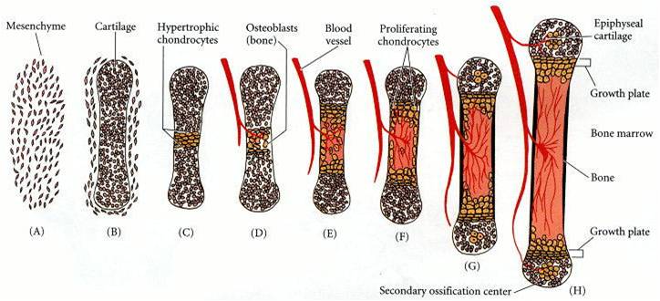
When babies are born, their skeletal system contains 300 components. These components are actually pieces of cartilage, which undergo a process called ossification, that allows them to become bone. The bony skeletal structure begins to develop at 8 weeks, endochondral ossification begins at the second month, and bone growth continues even after birth.

Bony skeleton development: 


Cartilage contains no blood vessels or nerves, and are surrounded by perichondrium, which prevents outward growth. Some of the cartilage remains, however, such as that found in the nose and ear. Cartilage can be classified as hyaline, elastic, or fibrocartilage. Matrix is secreted both inside and against the outside of the bone, through appositional and interstitial processes. This calcifies, or hardens the bone. Cartilage calcifies during normal bone growth, as well as during old age, which causes arthritis.

The pieces that do ossify often fuse together, creating a skeletal system of 206 bones by the end of development. Bones can be classified into two categories: axial skeleton and appendicular skeleton. The axial skeleton contains the bones of the skull, vertebral column, and rib cage. The appendicular skeleton contains the bones of the upper and lower limbs, as well as the shoulders and hips. Bones can also be classified as long bones, short bones, flat bones, and irregular bones. Long bone growth generally stops around the end of puberty, which prevents you from getting taller.

Fetal skull before fusion:


Skull after fusion:

The Ossification Process 
There are two types of ossification: intramembranous ossification, which replaces the thin connective tissue membrane with bone, and endochondral ossification, which replaces fetal cartilage with bone. Both types, however, rely on the thyroid hormone calcitonin for the regulation of calcium metabolism.

In bone production, there are three really important cells to consider: osteoblasts, osteoclasts, and osteocytes. Since your bones are always either growing or adapting to support your body, there are cells that will actually destroy bone, and others that will repair the bone to support its new requirements. Osteoclasts originate in bone marrow, and move to the bone surface where they will eat away old bone or protuberances that are out of place. Osteoblasts also come from the marrow and make their way to the surface to fill in cavities and restore bone. After their job is done, they flatten and line the surface of the bone. They then work to regulate calcium, as well as activate osteoclasts through the production of special proteins. Osteocytes come from osteoblasts and can be found inside the bone. As the osteoblasts build the bone, some surround themselves with new material while extending "arms", called dendrites, to other osteocytes. Osteocytes sense pressures and cracks, and help direct osteoclasts to where the bone needs to be dissolved.

The process of ossification:


Diagram of osteoclasts, osteoblasts, and osteocytes in action:


Diagram depicting bone growth and remodeling:


And there you have it: the background of bone growth, development, and remodeling!


Tuesday, November 3, 2015

Integumentary System

In this blog post, we are going to discuss the normal structure of skin and how certain conditions can change the normal structure and functions of the skin. Specifically, we are going to look at tattoos, laser hair removal, and poison ivy. Before we get into that, however, we should take a look at how skin functions under normal conditions.

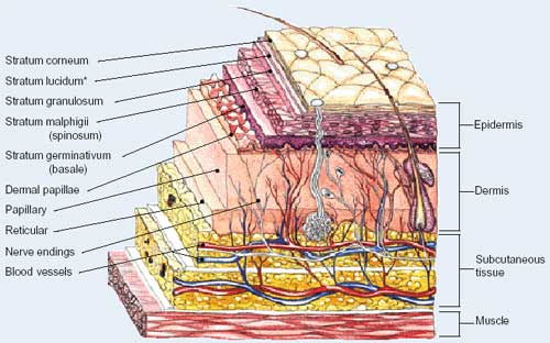
The skin consists of three basic layers: the epidermis, dermis, and hypodermis. Within these layers, there are other layers as well. The epidermis is composed of five layers: stratum basale (basale layer), stratum spinosum (prickly layer), stratum granulosum (granular layer), stratum lucidum (clear layer), and stratum corneum (horny layer). These five layers contain four distinct cell types, including melanocytes, Merkel cells, Langerhan's cells, and keratinocytes. The dermis consists of two layers: the papillary and reticular layers. There are other components to the skin as well, such as sweat glands, blood vessels, and hair follicles. The skin layers, under normal conditions, are shown below.


The picture below illustrates the process of dying the skin through tattoos. The needle pierces the epidermis and injects ink into the dermis in the papillary layer. The needle is motorized, puncturing the skin between 50 and 3,000 times per minute, depositing ink with every puncture. The ink can then be seen through the epidermis, and due to the stability of the cells in the dermis, the ink does not travel or fade in the skin. 


In the next picture, the integumentary system's reaction to poison ivy is displayed. An irritant from the ivy is transferred to the epidermis where it immediately triggers a reaction. Effector T cells signal white blood cells to attack the site of intrusion. A water soluble, nonallergenic compound is then formed, which prevents further allergic response. The Effector T cells then stop signaling the white blood cells, and natural antiseptic properties sanitize the rash, which is healed within 3 days. 


The last condition we are going to look at is electrolysis, or laser hair removal. In this case, a needle is inserted into the skin to come in contact with a hair follicle. An electrical shock is transmitted to the base of the follicle, causing it to reach a temperature in which hair growth is irreversibly inhibited. The epidermis, located below, remains undamaged.

http://www.follikill.com/wp-content/uploads/2014/01/how-laser-hair-removal-work1.jpg


I hope you enjoyed learning about the integumentary system!