Monday, February 22, 2016
The Effects of Heroin on the Brain
As we dig farther into neurons and their physiology, we are beginning to look at how they function in the brain. While doing this, our class was assigned to select a research topic that would enhance our understanding of neuron functionality. For this assignment, I chose to research how heroin affects the brain. My presentation on this topic can be found here; I hope you enjoy it!
Monday, February 8, 2016
Neurophysiology
Neurophysiology is a very interesting subject that works with three main components: neurons, nervous systems, and nerves. Today, we are going to look at how they function, beginning with the anatomy of the neuron.
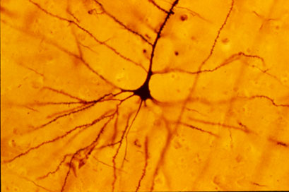
The neuron is a cell, and so it has many common components such as a nucleus, cell membrane, cell body, and different channels that ions can travel through. The neuron works to communicate signals to other neurons, muscles, and even glands. When they communicate, the signal they send causes the muscle to contact, the gland to secrete, or the next neuron to continue passing the signal through the nervous system. Neurons come in many shapes and sizes, however, all neurons have dendrites and one axon that extends from the cell body, a receptive portion, and a signaling portion.
The axon acts as the conducting or transmitting portion of the neuron, passing the signal along to the next neuron through the axon terminal and synapse. The axon connects to the cell body through the axon hillock. Axons can be from 1-2 inches to longer than a meter.
The cell body acts as a communicator between the dendrites and the axon. As the signal is received through the dendrites, the cell body passes it along to the axon. The cell body is also the main nutritional and metabolic portion of the cell.
The dendrites act as the cell receptors, receiving signals from other neurons and passing those signals through the cell. The flow of energy is directional in neurons, flowing from the dendrites, through the cell body and axon, into the next cell.
Some axons are covered in insulation bodies called myelin sheaths. These sheathes are made when Schwann cells wrap around the axon, releasing cytoplasm as it compresses in a spiral-like structure. The spaces between these sheaths are known as nodes of Ranvier. These nodes occur between every sheath, and allow ions to flow across the membrane.

There are four main channel types within the nervous system: the Sodium leak, Voltage Gated Na+, K+ Potassium Leak, and Voltage-Gated K+ channels. These ion channels are embedded in the neuron membrane, and work to control the movement of ions across the membrane. They are selective, regionally located, functionally unique, and either passive or active. Without these channels, there would be no possibility of neuron excitability.
Ions channels are selective, in that the ions they permit to cross the membrane must fit a certain criteria. The ion must have a certain size, charge, and must attract and hold a certain level of water. These standards vary between the different channel types.
There are also active and passive channel types. Active channels open and close, while passive channels are always open and allow ions to flow freely across the membrane.
Some active channels have gates that are controlled by voltage; membranes with more positively concentrated ions on the outside and more negatively concentrated on the inside create membrane potential, or resting potential, producing a voltage that opens the channel. Channels can also be opened by chemical processes. Neurotransmitters can bind to certain active channels and force them open, allowing ions to travel through.
Different ion channel types can be found on different parts of the neuron. Passive channels can be found on the dendrites, cell body, and axon; chemically gated channels can be found on the dendrites and cell body; voltage gated channels can be found on the axon hillock, unmyelinated axons, and the nodes of Ranvier.

The Sodium/Potassium pump works to keep a balance of sodium and potassium inside and outside of the cell. The pump works with an active process involving ATP hydrolysis, transforming ATP into ADP+P, which allows potassium to enter the cell and forces sodium out.

Resting potential, or RP, occurs when the inside of a neuron is more negative (-70mV) than outside of the neuron membrane. No change in voltage will occur over time, and the sodium and potassium will not reach equilibrium, as the outside of the neuron will have more sodium and the inside will have more potassium.

Synaptic potentials occur at -55mV, when a receptor on the dendrite receives a signal sent by the axon terminal. Such signals are referred to as neurotransmitters. When neurotransmitters are received, other molecules are permitted to pass through, such as Na+. mV will increase for a short time, while this potential is active, and then return to normal when it is no longer needed.

Action potentials occur during a brief reversal of membrane polarization, in which mV increases suddenly and greatly (-70mV to 30mV). In this type of situation, Na+ is allowed to travel into the membrane, and K+ is forced out. This potential only occurs in the axon, and is commonly known as a nerve impulse.

Thanks to nerves and their amazing communications, thoughts like this:

can turn into actions, like this:

Subscribe to:
Comments (Atom)