Wednesday, October 7, 2015

Human Models of Epithelia


Epithelial tissues are composed almost entirely of cells, and are supported by connective tissue. Within epithelial tissues, there are orginizational categories including: simple and stratified. Within those categories, there are also three types: squamous, cuboidal, and columnar.  In this post, our classmates worked together to create human models of the aforementioned epithelial tissues. 

The first model we made was for pseudostratified columnar epithelial tissue. This is a single layer of cells, in which all of the cells are of varying heights. Some cells do not reach the surface, and the nuclei can be seen on different levels. This type of epithelial tissue secretes mucus and propels it through ciliary action. Pseudostratified columnar epithelial tissue can be found lining the trachea and most of the upper respiratory tract. Nonciliated forms of this tissue can be found in male's super-carrying ducts and ducts of large glands.


Transitional epithelial tissue consist of many cell layers in which the cells located at the base are cuboidal and the surface cells are shaped like domes. Translational epithelia can be found lining the urinary bladder, ureters, and part of the urethra, stretching to permit the distension of the urinary bladder. 

The simple squamous is a stout, squished layer of single cells that diffuses and filters. It is used in the lymphatic and cardiovascular systems, creating a thin, slick layer that reduces friction. 


Stratified cuboidal is more uncommon in the body, and is usually 2-3 layers thick. Stratified cuboidal tissue can be found in some mammary and sweat glands.


Simple cuboidal epithelia is another example of epithelia that absorbs and secretes. This type of epithelia is found in the kidney tubules, ovary surface, and ducts and secretory portions of small glands. Simple cuboidal epithelial tissue is composed of a single layer of cube like cells with large, spherical central nuclei. 


Simple columnar epithelia consist of a single layer of tall cells with oval nuclei. Many of these cells contain cilia, which help transport mucus. This layer may also contain mucus-secreting unicellular glands referred to as goblet cells. Located in the digestive tract, gallbladder, and excretory ducts of some glands, nonciliated simple columnar epithelia works to absorb, secrete mucus, enzymes, and other substances. Ciliated simple columnar epithelia is found lining small bronchi, uterine tubes, and some regions of the uterus, working to propel mucus.

Stratified columnar tissue is also not very abundant in the body, however, it can be found in the pharynx, male urethra, lining of some glandular ducts, and transitional areas between 2 types of epithelia.


And thus concludes the lesson on epithelia! Thanks to my classmates and their efforts in making this lesson!

Histology Microscope Lab


When discussing tissues, there are four broad categories: epithelial, connective, muscle, and nerve. For this post, we are going to use a microscope to view these types of tissue on a much greater scale, describing each as we work. All of the photos were taken during the lab.

The first sample we observed was that of hyaline cartilage, which is classified as a connective tissue. When functioning, hyaline cartilage supports, reinforces, cushions, and resists compressive stress.
Hyaline cartilage is composed of a network of collagen fibers that have formed an imperceptible network and chondroblasts that produce the matrix.
   

The next sample we worked with was bone, also a type of connective tissue. Bone is comprised of hard, calcified matrix that contains many collagen fibers.    Bone works to support and protect while providing leverage for the muscles. In addition, bone stores calcium, minerals, and fat, and bone marrow is home to cell formation.


Next, we looked at smooth muscle tissue. Spindle-shapes cells make up this tissue, arranging themselves closely to form sheets that propel substances or objects along passageways without voluntary control.


We also looked at cardiac muscle. When in action, contracting tissue propels blood into circulation, also without voluntary control. This tissue is composed of branching, striated, and generally uninucleate cells that interdigital with at specialized junctions.


This next sample is taken from an involuntary muscle, meaning that it originates either in the cardiac system, or as a smooth muscle tissue in the walls of hollow organs. When compared to cardiac tissue, it does not share as many characteristics as it does when compared to smooth muscle tissue. It appears to have closely arranged cells with central nuclei, as do the cells found in smooth muscle tissue.


The next two samples are those of nerve endings and neuron motors.  These are composed of branching cells, with the cell processes extended from the nucleus-containing cell body. These transmit electrical signals, controlling activity in the body.

Monday, October 5, 2015

Homeostasis Lab


In a previous post, we discussed homeostasis and how it functions. The next question to ask is: how can we prove homeostasis occurs with changes to our internal/external environment? To answer this question, our group designed a lab in which our blood glucose levels were measured as we altered our diets.

For the first part of our lab, each member in our group fasted, eating nothing and drinking only water for 24 hours. We measured our blood glucose levels before, during, and after the fast. Considering the aspects of the experiment, it was hypothesized that our blood glucose levels would decrease. This was then supported through the experimental results, which are illustrated below. 


As you can see, the blood glucose levels decreased during the 24 hours. In the first graph there is an increase in glucose levels after the 24 hours, which occurred due to the fact that the subject took their blood after eating when the fast ended, where the other subjects did not.These readings were very interesting, as a hypoglycemic and vegan were tested within the experiment. Overall, this experiment was successful in proving homeostasis.


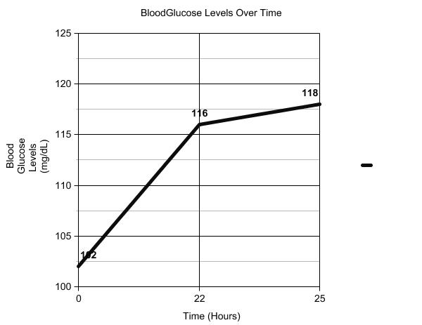
The second part of the lab involved one member of our group eating nothing but sugar-based foods for 24 hours, again measuring their blood glucose levels before, during, and after the 24 hours. For this part of the experiment, it was hypothesized that the blood glucose levels would increase over the 24 hours. The results are shown below.


As you can see, the glucose levels increased over the 24 hours, from 102 mg/dL to 118 mg/dL, again successfully proving homeostasis.